Database Open Access
Two-tiered response of cardiorespiratory-cerebrovascular networks to orthostatic challenge
Andras Eke , Peter Mukli , Orestis Stylianou , Andras Hartmann
Published: May 17, 2021. Version: 1.0
When using this resource, please cite:
(show more options)
Eke, A., Mukli, P., Stylianou, O., & Hartmann, A. (2021). Two-tiered response of cardiorespiratory-cerebrovascular networks to orthostatic challenge (version 1.0). PhysioNet. https://doi.org/10.13026/73w7-x881.
Please include the standard citation for PhysioNet:
(show more options)
Goldberger, A., Amaral, L., Glass, L., Hausdorff, J., Ivanov, P. C., Mark, R., ... & Stanley, H. E. (2000). PhysioBank, PhysioToolkit, and PhysioNet: Components of a new research resource for complex physiologic signals. Circulation [Online]. 101 (23), pp. e215–e220.
Abstract
The authors used this dataset to evaluate the responsiveness of the cardiorespiratory-cerebrovascular networks by capturing linear and nonlinear interdependencies to postural changes. Ten young healthy adults participated in our study between December 2009 and February 2010 at Semmelweis University, Faculty of Medicine. Non-invasive measurements of arterial blood pressure (ABP), cardiac cycle duration (derived from ABP signals), breath-to-breath interval (capnography), cerebral blood flow velocity (BFV, recorded by transcranial Doppler sonography), and cerebral hemodynamics (hemoglobin signals monitored by near-infrared spectroscopy) were performed for 30-minutes in resting state, followed by a one-minute stand-up and a one-minute sit-down period. BFV and ABP signals were calibrated, and the dataset contains values in cm/s and mmHg, respectively. NIRS measurement yielded intensity values (raw signals) from which change of HbO (oxyhemoglobin), HbR (deoxyhemoglobin) and HbT (total hemoglobin) concentration in tissue can be calculated (in micromol/liter). Raw NIRS signals were synchronized to BFV, ABP and capnography time series yielding 33 minutes of resampled data (sampling frequency: ~ 3 Hz).
Background
Dynamic interdependencies within and between physiological systems and subsystems are essential for the organism to maintain its steady-state via a plethora of homeostatic mechanisms [1]. The aim of physiological regulation is to restore the optimal state of the internal environment that is challenged by continuous perturbations. The deviation of actual value from its physiological set point elicits multiple negative feedback loops that occur at various time scales resulting in a delayed response with an amplitude proportional to perturbation [2-4]. The recently introduced concept of Network Physiology offers a novel framework for an enhanced characterization, quantification, and understanding of the dynamical interactions between organ systems underlying homeostatic adaptation [5-7].
These interactions mediate regulatory responses elicited by various perturbations, such as the high-pressure baroreflex and cerebral autoregulation, alleviating the impact of orthostatic stress on cerebral hemodynamics and oxygenation [8]. Under physiological conditions, high-pressure baroreflex helps to adapt to orthostatic stress - a consequence of an abrupt change in body position promptly altering the central arterial blood pressure (ABP) - while cerebral blood flow is maintained by autoregulatory mechanisms within a range of ABP values. The dataset was collected as part of a study to evaluate the responsiveness of the cardiorespiratory-cerebrovascular networks by capturing linear and nonlinear interdependencies to postural changes.
Methods
A total of 10 healthy young adults - students and employees of Semmelweis University - were recruited for participation in this study (age: 26.3±3.7 years, 5-5 female and male). None of them reported neurological, psychiatric or cardiovascular diseases or living with the condition of orthostatic hypotension. One male subject was excluded due to a lack of acoustic window necessary for transcranial Doppler (TCD) measurements (see below). Written informed consent was obtained from all subjects prior to participation.
The measurement protocol consisted of a 30-minutes resting-state period followed by a one-minute stand-up and one-minute sit-down period. Subjects were asked to change body position abruptly. The protocol was approved by Regional and Institutional Committee of Science and Research Ethics (ethical approval number 53/2009).
Arterial blood pressure was recorded non-invasively by using tonometric monitors (Colin BP-508, Colin Medical Technology, Corporation, Komaki City, Japan). The array of transducers - embedded in a wristband adjusted to the participant - captured signals from the right radial artery over the styloid process. Capnographic measurements were carried out with the same device. Blood flow velocities were recorded bilaterally in the middle cerebral arteries with the aid of 2 MHz transducers in pulsed-wave mode (DWL Multidop-T, Sipplingen, Germany). Transducers fitted in a probe holder mounted on the head of the participant that ensured their position during measurement. Signals were digitized at a 100 Hz sampling rate with DT9816 data acquisition device (Data Translations, Marlborough, Massachusetts, United States) and were captured by Winview LE software (Team Solutions Inc., Grande Vista Ave Laguna Niguel, California, United States).
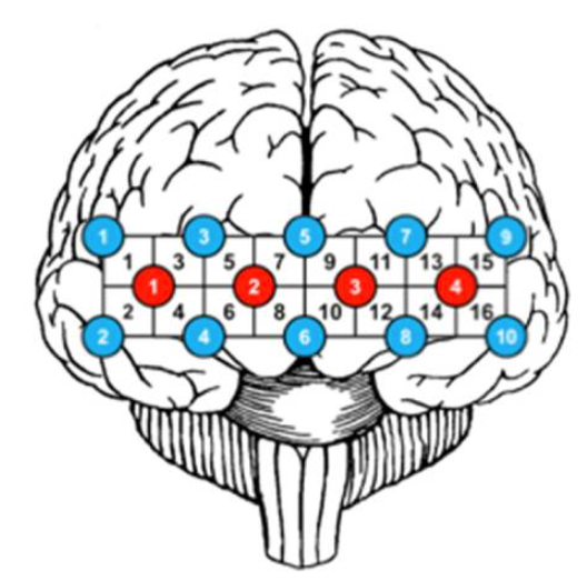
The consecutive values of cardiac cycle durations (an estimate of RR-interval) were extracted from blood pressures using the algorithm described in [9-10]. From the respiratory record, we created the breath-to-breath interval time series (BB) were reconstructed from the respiratory signal with the aid of the peakfinder function of MATLAB (version 2012, Mathworks, Natick, MA, USA). All light intensity signals were recorded from 16 regions of the prefrontal cortex at the following wavelengths: 730 nm, 805 nm and 850 nm. Finally, TCD, CCD, and BB time series were resampled using timestamps corresponding to NIRS signals and markers set during the measurement protocol (resample function of MATLAB). All signals (including BB and RR) were resampled at 3 Hz (interp1 function of MATLAB). The method of resampling was a first-order linear interpolation.
Data Description
The uploaded data contains the signals resampled with respect to the same sampling time. Data for each subject is stored in a separate file. The CSV files contain all measured data resampled to 3 Hz. Subject 05 was excluded due to an inadequate transtemporal window.
- Column 1: TCD signal from a right medial cerebral artery (calibrated)
- Column 2: TCD signal from a left medial cerebral artery (calibrated)
- Column 3: Arterial blood pressure signal (calibrated)
- Column 4: Respiratory signal (uncalibrated)
- Column 5-52: Intensity values measured by near-infrared spectroscopy (NIRS) from channel 1-16
- Columns 5, 8, 11, 14, 17, 20, 23, 26, 29, 32, 35, 38, 41, 44, 47, 50: λ=850 nm
- Columns 6, 9, 12, 15, 18, 21, 24, 27, 30, 33, 36, 39, 42, 45, 48, 51: λ=730 nm
- Columns 7, 10, 13, 16, 19, 22, 25, 28, 31, 34, 37, 40, 43, 46, 49, 52: λ=805 nm
- Column 53: Time stamp
Collection of chromofor concentration values started after calibration of NIRS, ~37 seconds (first four markers). Markers were set according to protocol and in case of artefacts. Regularly we set the following markers:
- ~1800s – end of resting period, preparing for maneuver
- ~1860s – participant stands up promptly
- ~1920s – participant sits down promptly
- ~1980s – end of sit-down period
Further details are provided in the data_info file. Channel layout is displayed in channel_layout.png, reproduced from Figure 1 of Racz et al. [11].
Usage Notes
Data are published as CSV files which can be directly imported into common data analysis software. The data is not preprocessed therefore it is necessary to remove motion artefacts. To eliminate these from NIRS signals, we recommend a wavelet filtering method [12]. Signal component of interest can be enhanced by using Butterworth filters in the frequency domain [13] and the correlation based signal improvement algorithm [14].
With the aid of this dataset, cerebral autoregulation, baroreflex function and spontaneous neurovascular coupling can be studied. The simultaneously recorded TCD, BP, NIRS and BB signals support elimination of systemic components from cerebral hemodynamic fluctuations. Findings from this dataset have been published in the paper of Mukli et colleagues [15] entitled "Two-Tiered Response of Cardiorespiratory-Cerebrovascular Network to Orthostatic Challenge". In this study, a network physiology approach was implemented using a linear and model-free estimator to characterize the effects of postural changes on the interdependent physiological processes in different frequency bands.
Limitations
Since the stand-up and sit-down maneuvers were inherently associated with perturbations generating large and transient motion artifacts in the physiological records, it was necessary to exclude the very early phase of the postural challenge from our analysis [15]. Consequently, assessing dynamic cerebral autoregulation is difficult to do reliably. The time periods after the resting-state may be too short for certain stochastic time series analysis due to a limited number of data points necessary for statistical reliability. Recovery of hemodynamics cannot be fully assessed in the last sit-down period since one minute was found to be too short to achieve a new steady-state.
Acknowledgements
We would like to thank all participants of the study. We acknowledge the support from
- Prof Britton Chance who designed and constructed (NIM Inc., University of Pennsylvania, USA) the near-infrared imager used in the study
- Semmelweis University, Department of Neurology for providing the device for transcranial Doppler sonography (DWL Multidop-T, Sipplingen, Germany) and blood pressure measurement (Colin BP-508, Colin Medical Technology Corporation, Komaki City, Japan).
Conflicts of Interest
The authors declare no conflict of interest.
References
- Cannon WB. ORGANIZATION FOR PHYSIOLOGICAL HOMEOSTASIS. Physiol Rev. 1929;9(3):399-431.
- Ivanov PC, Nunes Amaral LA, Goldberger AL, Stanley HE. Stochastic feedback and the regulation of biological rhythms. Europhysics Letters. 1998;43(4):363-8.
- Ashkenazy Y, M. Hausdorff J, Ch. Ivanov P, Eugene Stanley H. A stochastic model of human gait dynamics. Physica A. 2002;316(1):662-70.
- Lo CC, Amaral LAN, Havlin S, Ivanov PC, Penzel T, Peter JH, et al. Dynamics of sleep-wake transitions during sleep. Europhysics Letters. 2002;57(5):625-31.
- Bashan A, Bartsch RP, Kantelhardt JW, Havlin S, Ivanov P. Network physiology reveals relations between network topology and physiological function. Nat Commun. 2012;3:702.
- Bartsch RP, Liu KKL, Bashan A, Ivanov PC. Network Physiology: How Organ Systems Dynamically Interact. PloS one. 2015;10(11):e0142143.
- Lin A, Liu KKL, Bartsch RP, Ivanov PC. Dynamic network interactions among distinct brain rhythms as a hallmark of physiologic state and function. Commun Biol. 2020;3(1):197.
- Koeppen, and Bruce A. Stanton. Berne & Levy Physiology. Philadelphia, PA: Mosby/Elsevier, 2010.
- Pan J, Tompkins WJ. A real-time QRS detection algorithm. IEEE transactions on biomedical engineering. 1985(3):230-6.
- Sun J, Reisner A, Mark R, editors. A signal abnormality index for arterial blood pressure waveforms. 2006 Computers in Cardiology; 2006: IEEE.
- Racz FS, Mukli P, Nagy Z, Eke A. Increased prefrontal cortex connectivity during cognitive challenge assessed by fNIRS imaging. Biomedical Optics Express. 2017;8(8):3842-55.
- Molavi B, Dumont GA. Wavelet-based motion artifact removal for functional near-infrared spectroscopy. Physiological measurement. 2012;33(2):259-70.
- Kirilina E, Jelzow A, Heine A, Niessing M, Wabnitz H, Bruhl R, et al. The physiological origin of task-evoked systemic artefacts in functional near infrared spectroscopy. Neuroimage. 2012;61(1):70-81.
- Cui X, Bray S, Reiss AL. Functional near infrared spectroscopy (NIRS) signal improvement based on negative correlation between oxygenated and deoxygenated hemoglobin dynamics. Neuroimage. 2010;49(4):3039-46.
- Mukli P, Nagy Z, Racz FS, Portoro I, Hartmann A, Stylianou O, et al. Two-Tiered Response of Cardiorespiratory-Cerebrovascular Network to Orthostatic Challenge. Frontiers in Physiology. 2021;12(216).
Access
Access Policy:
Anyone can access the files, as long as they conform to the terms of the specified license.
License (for files):
PhysioNet Contributor Review Health Data License 1.5.0
Data Use Agreement:
PhysioNet Contributor Review Health Data Use Agreement 1.5.0
Discovery
DOI (version 1.0):
https://doi.org/10.13026/73w7-x881
DOI (latest version):
https://doi.org/10.13026/ehkk-8y70
Corresponding Author
Files
Total uncompressed size: 17.8 MB.
Access the files
- Download the ZIP file (6.6 MB)
-
Download the files using your terminal:
wget -r -N -c -np https://physionet.org/files/cardioresp-response-orthostat/1.0/
-
Download the files using AWS command line tools:
aws s3 sync s3://physionet-open/cardioresp-response-orthostat/1.0/ DESTINATION